

Vertebral column
The vertebral column supports the head and body, serves as an attachment point for many muscles, and protects the delicate spinal cord. Here we’ll cover all the segments, processes, foramina, and articulations of the vertebral column itself!
The vertebral column
We've all heard or are familiar with old phrases like "put your back into it" or "stand up straight" or perhaps we've considered someone "spineless" when they didn't rise to a challenge. Well, the fact that the vertebral column or euphemisms for it are all around us underlies how important these structures are to our life and well-being.
The vertebral column is made of a series of bony vertebrae, the cartilaginous discs between them, and a myriad collection of ligaments to reinforce the stack of bones and cartilage. Let's review these elements and familiarize ourselves more with "the spine."
Functions of the vertebral column
Its appearance in metaphors aside, the vertebral column has several anatomical and physiological functions in the body. Primarily it serves to protect the spinal cord, support the skull, and serve as an attachment point for ribs, many muscles, and lots of ligaments.

This stack of bone must be strong yet flexible to accommodate all the movements, positions, strength, weight-bearing, and support that we ask of it. Each element and each segment of the vertebral column is specialized to allow us to walk, run, dance, turn, bend, sit up straight, or whatever else we need to do.
Segments of the vertebral column
I'm not one to memorize the numbers of bones in any given part of the body, but once we work with them a few times, it will become easy to count up that there are 33 vertebrae total, all working to support the spinal cord, skull, and the many muscle and connective tissue structures attached to it.
There are several segments within the vertebral column and this page describes cervical, thoracic, lumbar, sacral, and coccygeal segments, along with the ligaments that support them.

When they speak of "the spine," most people mean those regions found in the neck, mid-back, and lower back. Those are collectively known as the "presacral" section, or those portions above the sacrum and coccyx. Of the 33 total elements of the vertebral column, 24 of them are presacral -
7 cervical, 12 thoracic, and 5 lumbar vertebrae.
Each segment gets a section below, but before we get to the peculiarities of each region and some special members of each segment, let's review the features that most vertebrae have in common.

Features common to most vertebrae
Each vertebra shares some features with the others (well, most of them...a few are very peculiar and those get their own treatment below). Let's review common features of most vertebrae:
Vertebral body is the anterior portion that supports the weight of the rest of the body
Vertebral arch is everything posterior to the vertebral body, consisting of the pedicles, lamina, and spinous process; the arch protects the spinal cord
Pedicles are two “feet” that connect both lamina to the vertebral body
Lamina are two flat processes that connect pedicles to spinous process
Vertebral foramen is the opening between the body and arch
Vertebral canal is created when vertebrae are stacked up (they are continuous vertebral foramina); the spinal cord, meninges, roots of spinal nerves, and vasculature are all within the vertebral canal
Spinous process is one midline posterior projection formed where the two laminae meet - this is the bump of bone visible and palpable along the middle of the back
Transverse processes (there are two) project laterally from the junction of pedicles and laminae

Articular processes (there are four) occur on most vertebrae - two project superiorly, and two project inferiorly from the point near where the pedicles and laminae meet
Articular facets (zygopophyseal joints) are on articular processes, allowing the vertebrae to stack up on one another; there are two superior articular facets (they articulate with the inferior articular facets of the vertebrae above) and two inferior articular facets (they articulate with the superior articular facets of the vertebrae below)
Vertebral notches are indentations near the junction of the pedicles and body, best seen on the lateral aspect; the superior vertebral notch lies between the body and superior articular process, the inferior vertebral notch is an indentation of the pedicle
Intervertebral foramen are formed between each superior and inferior vertebral notch when the vertebrae are stacked - these contain the dorsal and ventral nerve roots forming a spinal nerve, and often the dorsal root ganglion
Vertebral curvatures
The vertebral column has a few normal curvatures, formed both at birth and as we learn to walk.
Primary curvatures (x2) are the ones present at birth. These are concave anteriorly - called kyphotic curves. They are found naturally in the thoracic and sacral segments of the vertebral column.
Secondary curvatures (x2) develop as one learns to walk. These are convex anteriorly - called lordotic curves. These are found naturally in the cervical and lumbar regions.
Abnormal curvatures of the vertebral column may develop, too. Exaggerated kyphosis of the thoracic segment is most common - this is called hyperkyphosis. The lumbar region may also become exaggerated in curvature, called hyperlordosis. Finally, abnormal lateral curvatures to either side are called scoliosis.


Cervical vertebrae
There are 7 total cervical vertebrae so they are numbered 1 through 7. The first two cervical vertebrae are quite unique so they'll get special treatment below. First, let's cover the typical cervical vertebrae.
Typical cervical vertebral bodies are small and oval shaped. The bodies have little buildups of bone on the lateral edges called uncinate processes. The small, oval body is a feature unique to cervical vertebrae - they support the head but not as much weight as the rest of the vertebral column.
To facilitate rotation of the head and neck, articular facets in the cervical region are oriented in the transverse plane.
Another unique feature of cervical vertebrae are bifid spinous processes - these have two projections posteriorly. If you see a bifid spinous process, you must be looking at a cervical vertebrae.
Transverse processes of cervical vertebrae have short anterior and posterior tubercles with a groove between them. The groove on the transverse process contains the spinal nerve whereas the tubercles serve as attachment for muscles of the neck and back.

Yet another unique feature of cervical vertebrae are transverse foramen within the transverse processes. This is an important structure, as it allows the large vertebral artery and a few smaller vertebral veins to move through the neck in relation to the skull. The vertebral artery is a significant source of blood supply to the spinal cord, brain, and cranial cavity.
The vertebral artery, a branch of the subclavian artery in the root of the neck, enters the transverse foramen at the C6 level. It courses superiorly through the transverse foramen from C6 to C1. It then courses along the superior aspect of C1, forming a large groove there, then turns to ascend through the foramen magnum and into the cranial cavity. The vertebral vein descends the transverse foramen from C1 to C7 levels (the artery is not in the C7 level, just the vein).
Atlas - first cervical vertebrae
The first cervical vertebrae is quite unique and deserves its own section. Because it supports the skull directly, C1 is also known as the atlas. It has several unique characteristics:
It is the widest cervical vertebra, to help support the skull
The superior aspect of the arch has a wide groove for the vertebral artery
C1 has no true spinous process OR body; instead, it has an anterior and posterior tubercle on arches that connect to the lateral mass to form a ring of bone
The contour of C1's superior articular facets (on the lateral mass) are notched because they articulate with occipital condyles of the skull (inferior articular processes and facets are more oval, as in the rest of the cervical vertebrae)
Though it has no body or spinous process, C1 does have typical transverse foramina on the transverse processes

The axis - second cervical vertebrae
The 2nd cervical vertebra is also unique. Because of its function in allowing side-to-side rotation of the head it is also known as the axis. Like the atlas, the axis has a few unique characteristics, and a few features similar to the rest of the cervical vertebrae.
It is the strongest cervical vertebra with a stout, wide, and tall vertebral body
The body of C2 has a superior projection, the dens or odontoid
process, which allows rotation of the skull by articulating with C1 (it leaves a facet inside of anterior arch of C1, in fact).
The spinous process is bifid, and prominently so since several upper neck muscles attach to it.
The transverse processes have transverse foramina like the rest of the cervical vertebrae

Thoracic vertebrae
Thoracic vertebrae are found in the upper and middle back regions. They are numbered 1 through 12. Thoracic vertebrae share several characteristics unique to this region.
Vertebral bodies are heart-shaped and intermediate in size between the cervical and lumbar regions. They bear more weight than the cervical bodies but not as much as the lumbar ones.
Spinous processes of thoracic vertebrae are quite long, triangular in shape, and point obliquely inferior.
To facilitate lateral bending and flexion of the back, thoracic vertebral articular facets are oriented in the coronal plane.

The defining and unique aspect of thoracic vertebrae is that they articulate with ribs at costal facets. Each thoracic vertebrae has one or more indentations on the vertebral body for articulation with the head of a rib. Most thoracic vertebrae articulate with two ribs because the head of a rib nestles between two thoracic vertebrae. These create "half" facets known as demifacets. The superior demifacet is for the rib head of the same level (T4 rib head with the T4 vertebral body). The inferior costal facet is for the rib below (so T5 rib head if we're still talking about the T4 vertebral level).
In a few cases, though, only one rib articulates with the body. For instance, T1 has just one full costal facet for articulation with rib 1. Then, T9 has only one costal demifacet on the body, for articulation with the head of the 9th rib. Similarly, T10 has just one full costal facet on its body, for the head of the 10th rib. Both T9 and T10 have transverse costal facets for articulation with the tubercles of ribs 9 and 10 respectively. Then, T11 and T12 each have one costal facet on their bodies (for rib 11 and 12, respectively) but their transverse processes do not have costal facets because those ribs do not have tubercles.
Lumbar vertebrae
Lumbar vertebrae are found in the lower back. They are numbered 1 through 5. They are very similar to one another and share these features:
Lumbar vertebral bodies are large - they bear the weight of all the vertebrae and body above them
Spinous processes are short, thick, and project posteriorly
Their articular facets are oriented closer to the sagittal plane to facilitate flexion and extension of the lower back (as in bending at the waist)

There are a few unique features in the lumbar region.
Mammillary processes are located on the superior articular process, allowing attachment for multifidus and intertransversarii muscles that help stabilize vertebral joints
Accessory processes are located on the posterior surface of the base of transverse processes, allowing for attachment of intertransversarii muscles
Sacral vertebrae
Sacrum composed of the 5 fused vertebrae. The first one, SV1, articulates with LV5 at the superior articular process and facet, as well as the ilium of the pelvis through the auricular surface. The coccyx articulates with SV5.
Each side of the sacrum, anterior and posterior, has distinct features. The anterior (or ventral) surface faces the inside of the pelvic bowl. It is smooth and curved. The posterior surface faces the gluteal region and is rough and bumpy with muscle attachments.

Features of the anterior (pelvic) side include:
Sacral promontory – an anterior projection of the lip of SV1 vertebral body
Ala - the "wings" of the sacrum, lateral to the anterior sacral foramina
Anterior sacral foramina – four pairs of foramina that are similar to intervertebral foramina of the CV to LV regions, these transmit ventral (anterior) primary rami of spinal nerves
Features of the posterior (gluteal) side include:
Median sacral crest (x1) – fused spinous processes of sacral vertebrae
Medial (intermediate) sacral crests (x2) – between median sacral crest and lateral margins of the sacrum
Sacral cornua - inferior projections of the intermediate sacral crests, they lie on each side of the sacral hiatus
Sacral hiatus is the inferior opening of the sacral canal (the continuation of the vertebral canal from above)
Posterior sacral foramina – four pairs of foramina that transmit dorsal (posterior) primary rami of spinal nerves
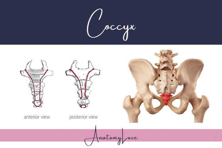
Coccygeal vertebrae
The coccyx is composed of fused coccygeal vertebral bodies numbering between 3 and 5 segments, usually 4. The first one articulates with the sacrum. The coccygeal vertebrae have an incomplete or absent vertebral arch. Though small, the coccyx does serve as an attachment point for muscles of the pelvic floor, and as an anchor for the filum terminale of the spinal cord.
Vertebral column wrap-up
Each vertebrae and each vertebral segment is unique. Taken together though, this stack of bones provides structural support, serves as attachments for many muscles, and protects important vessels and the spinal cord.
Check out other posts below - and follow AnatomyLove on socials. Thank you!