

Nasal cavity

Sniff, snot, snort - the nasal cavity has it all! From the bony housing of the nasal cavity, to the mucous-producing factories of the epithelium and glands, to sinuses and where that snot drains, this post covers all things nasal cavity!
Nose and external features
There are a lot of descriptions and euphemisms about our noses - long or short, pointy or round, I'll try to be "on the nose" throughout this post, starting with external features! Let's not take these for granted since the nose is one of the entryways into our body.
The "top" of the nose is the bridge and dorsum of the nose. It extends from the pinched part between the eyes (left and right nasal bones make up this part) to the tip of the nose (also called the apex). Two ala extend on either side of the dorsum, covering the naris or nostril. The ala have small cartilages within them, keeping the nostrils open or even flared when muscles pull on those cartilages and skin.
One of my favorite anatomy terms is a vestibule and there is one just inside the nostrils, called the nasal vestibule. The reason I like this one so much is because it is a unique region of the nasal cavity with an epithelial lining and specific features found no where else in the nose. The nasal vestibule is lined by stratified squamous keratinized epithelium, just like the rest of our skin. This is because we can (and often do!) touch it quite easily. It needs to be a bit more protective than the deeper linings of the nose that we can't quite reach. The vestibule is also like skin in that it has hairs, technically called vibrissae, with associated sebaceous glands that coat the hair with sebum to help trap particles and dust before they get any deeper. Neat!

Zones of the respiratory system
We shouldn't take for granted that the nose and nasal cavity is an entryway into the rest of the body - especially the respiratory system. The nasal cavity is the start of the conducting portions of the respiratory system - the part that accepts incoming air, conditions it, and funnels it to the lungs. The oral cavity, pharynx, larynx, trachea, and main bronchi complete the conducting portions of the respiratory system. Actual gas exchange, or respiration, happens in the respiratory portions inside the lungs themselves at bronchioles, alveolar ducts and sacs, and finally alveoli.

Bony framework of the nasal cavity
Several bones contribute to the framework of the nasal cavity - they are, after all, what makes it a cavity or space in the head.
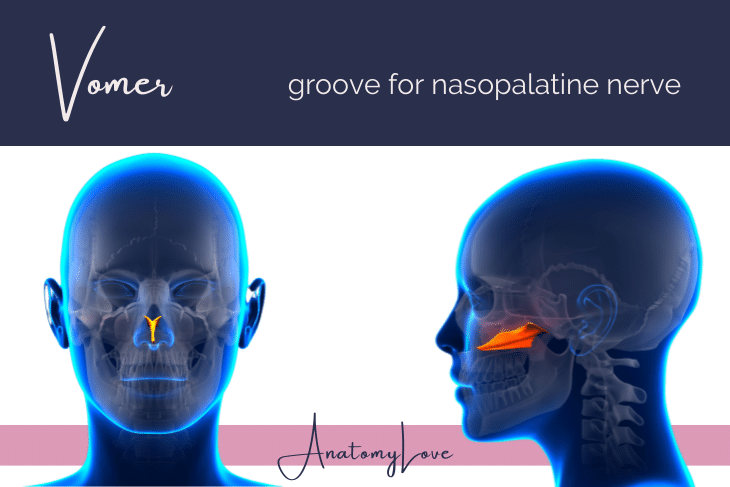
Let's start with the ones in the midline - the vomer and ethmoid bones. The vomer is a plate of triangle-shaped bone that forms the inferior/lower portion of the bony nasal septum. It has one distinguishing feature, a groove for the nasopalatine nerve.
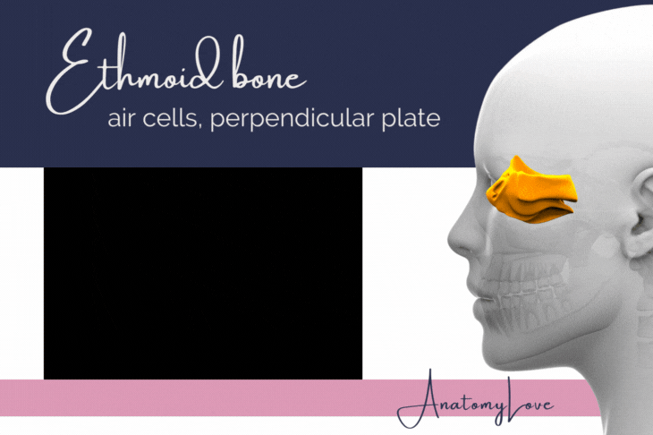
The ethmoid bone though, has a whole lot going on! Let's start right in the middle with the perpendicular plate of the ethmoid bone. It forms the superior/upper half of the bony nasal septum. Technically, the perpendicular plate has a sharp, bony point called the crista galli that projects into the anterior cranial fossa inside the cranial cavity itself. On either side of the perpendicular plate are spaces/sinuses called ethmoidal air cells. These cavities are numerous and quite varied, lined by respiratory epithelium (discussed below), and are part of the snot-making paranasal sinuses of the head (also discussed below).
Another important feature of the ethmoid bone, and the part for which it is named, is the cribriform plate. This thin plate of bone forms a roof over the air cells, separating the nasal cavity from the cranial cavity. The cribriform plate has many very small holes (foramina) that allow axons of olfactory neurons to reach the olfactory bulbs inside the cranial cavity. The word "ethmoid" derives from the Greek word "ethmos" or "ēthmoeidēs" meaning sieve. And that is certainly what it looks like with all those tiny holes!
We'll come back to more features of the ethmoid bone, the superior and middle nasal conchae that contribute to the lateral nasal wall, below.
Lateral nasal wall
The features of the bony nasal septum discussed above are right in the midline. Since the nasal cavity is a space, though, it also has bones that form the lateral nasal walls. The lateral walls of the nasal cavity have three bony projections, called conchae, spaces above and below the conchae, and many openings that allow the sinuses to drain. Let's run through them from superior to to inferior (top to bottom).

Nasal septum
The septum of the nasal cavity divides that space into left and right portions. The septum itself is made of bone and cartilage. The bony portions consist of the perpendicular plate of the ethmoid bone (posterosuperioly) and the vomer (posteroinferiorly). Septal cartilage (hyaline) fills in the anterior portions of the nasal septum.

Paranasal sinsues
These sinuses are the ones that produce "snot" and are typically what comes to mind when we think of head colds. The paranasal sinuses are spaces within bone that are lined by respiratory mucosa (pseudostratified ciliated columnar epithelium with goblet cells, supported by a connective tissue lamina propria that is rich in nerves and vessels). Let's run through the bones that include sinuses!
Frontal sinus is within the frontal bone entirely. These spaces are within the frontal bone, just above the eyebrows, especially towards the middle of the forehead. Each frontal sinus is unique! In the image of paranasal sinuses below the frontal sinus is green.
Sphenoidalsinus lies within the body of the sphenoid bone itself. The body and sphenoid sinus is located right in the middle of the head but just behind the orbits. The sphenoidal sinus is shown in purple on the image below.
Ethmoidal sinuses are also varied. Technically, there are three zones of ethmoidal sinuses - anterior, middle, and posterior. These are often referred to as the "ethmoidal air cells" because they are indeed filled with air (unless they are clogged with snot!) and are quite small. The ethmoid bone and these air cells specifically, lie right between the orbits. In the image below they are light blue.
Maxillary sinuses are the biggest spaces in the face. Shown in orange below, they lie completely within the maxillae bones, left and right. As the image shows, maxillary sinuses lie below/inferior to the orbits and lateral to the nose.

Drainage of nasal sinuses
I hope what comes next is perhaps why you came to this page - where do paranasal sinuses drain into the nose?! Let's run through them from superior to inferior, along the lateral nasal wall.
The sphenoethmoidal recess is an area superior to the superior nasal concha. It lies between the sphenoid bone and the ethmoid bone, thus the name. The recess is a small space that receives drainage from the sphenoid sinus.
The superior meatus lies just inferior to the superior concha. This space receives drainage from the posterior ethmoidal air cells. One way I help students remember this is to remind them that the superior concha itself is very posterior on the lateral nasal wall. The only thing that could open there are the posterior ethmoidal air cells.
The middle meatus has a whole lot going on! This space is located just inferior to the middle concha. This areas has many interesting bony features, too, like the semilunar hiatus, ethmoidal bulla, uncinate process of the ethmoid bone, and several openings for paranasal sinuses. First, the frontal sinus opens into the semilunar hiatus of the middle meatus via the frontonasal duct. The anterior ethmoidal air cells open into the frontonasal duct so they too drain into the semilunar hiatus and middle meatus. The middle ethmoidal air cells drain onto the ethmoidal bulla within the middle meatus. Finally, the large opening for the maxillary sinus lies within the semilunar hiatus, too. My mnemonic for keeping track of these four openings in the middle meatus is FAMM (as in, its lit, fam! lol) - which stands for Frontal, Anterior ethmoidal, Middle ethmoidal, and Maxillary sinuses.
Lastly, the inferior meatus receives the opening of the nasolacrimal duct which drains the lacrimal sac from the orbit. This sac is not a paranasal sinus since it drains lacrimal secretions from the eye, but it does open into the lateral wall of the nasal cavity so it deserves a quick mention!

Linings of the nasal cavity
Most of the nasal cavity is space within bone. But, that bone is not bare. It is covered with epithelial surfaces and mucosae that serve important functions in conditioning the air coming into our bodies, trapping particulates within that air, and allowing a sense of smell. The nasal cavity has two regions of mucosa that are specialized in what they do - let's take a look!
Olfactory mucosa
The olfactory mucosa lines the superior one-third of the nasal cavity, especially the posterior portions. This region includes special sensory neurons called olfactory receptors. These cells are surrounded by supporting cells and olfactory glands. The reason the olfactory region is superior within the nasal cavity is because axons leaving the olfactory neurons must travel superiorly, through the cribriform plate of the ethmoid bone, to reach the olfactory bulbs and tracts within the cranial cavity.

Respiratory mucosa
The rest of the nasal cavity is lined by respiratory mucosa, typical of what lines the conducting portions of the respiratory system (nose, larynx, trachea, bronchi). This lining consists of pseudostratified ciliated columnar epithelium with goblet cells - or just "respiratory epithelium," whew! Supporting that epithelium is a loose connective tissue lamina propria layer that is packed with nerves, arteries and veins, and seromucous glands. The respiratory mucosa helps warm and condition incoming air thanks to all those blood vessels. It also helps trap particulates in the air by secreting mucus from goblet cells and seromucous glands.

Pharyngeal tonsils
The entryways into the body (nose and mouth, if we're sticking to head-parts!) are important sentinels and protectors of the rest of the body. Lying at the back of the nose and mouth are special patches of lymphoid tissue we call tonsils. There are many clusters of these throughout the nasopharynx, oropharynx, and laryngopharynx.
The tonsils located at the back of the nose, technically embedded in the nasopharynx mucosa, are the pharyngeal tonsils, also known as "adenoids." This special patch of lymphoid tissue is right where a nasopharyngeal swab might try to reach when one must take a covid-19 test.
Lymphoid tissue in the form of tonsils are made of germinal centers packed with lymphocytes, ready to sense a harmful invader or pathogen and begin dividing to fight it off if necessary. The centers are embedded in the mucosa of their specific region. In this case, the pharyngeal mucosa is stratified squamous non-keratinized epithelium. Crypts, or clefts, in the epithelium allow increased surface area and contact with potentially harmful antigens and pathogens.

Neurovasculature of the nasal cavity
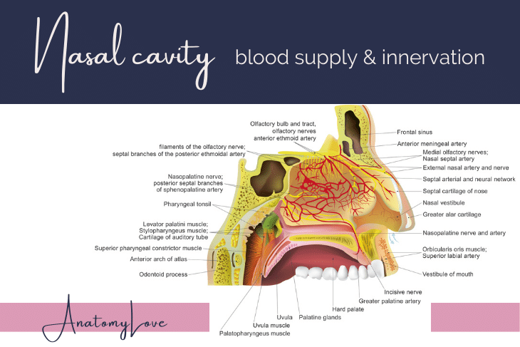
The nose and nasal cavity is rich with blood supply and innervations. The diagram below show a messy network of both arteries (and veins) as well as nerves. This is one reason the nose and nasal cavity are so sensitive and bleed easily.
Blood supply to the nasal cavity
The arteries that contribute to the nasal cavity are branches of the ophthalmic, maxillary, infraorbital, and facial arteries.
The ophthalmic artery (itself a branch of the internal carotid artery), courses through the orbit and forms the posterior and anterior ethmoidal arteries. Both of these supply mucosa of the ethmoidal air cells within the ethmoid bone, then move on to supply superior regions of the nasal cavity and septum. The anterior ethmoidal artery, especially, contributes not just to the nasal cavity but also skin over the dorsum of the nose,.
The third portion of the maxillary artery moves from the infratemporal fossa (deep face) to the pterygopalatine fossa, a bony space located posterolateral to the nasal cavity. Here, the third portion of the maxillary artery forms many branches. The one that supplies the nasal cavities is the sphenopalatine artery. This vessel courses from the pterygopalatine fossa through the sphenopalatine foramen, located on the superior and medial wall of the pterygopalatine fossa. Once in the nasal cavity, the sphenopalatine artery branches into several lateral branches to supply mucosa over the conchae and several septal branches.
The contribution of blood to the nasal cavity from the infraorbital artery is quite small, though still important. The infraorbital artery is itself a branch of the maxillary artery, just like sphenopalatine artery. However, the infraorbital supplies blood to the superficial face skin and muscles over the maxilla. It does form a lateral nasal branch that anastomoses with the facial artery to supply the alae of the nose and vestibule of the nasal cavity (where the nostrils are).
The facial artery is the main artery of the superficial face. As a branch of external carotid artery in the neck, the facial artery reaches the face by coursing over the inferior edge of the mandible. It forms several branches to the lips, cheeks, and nose. As it courses by the upper lip it forms a superior labial branch which sends a small artery into the anterior portions of the nose, too. The facial artery also forms lateral nasal branches which anastomose with branches of the infraorbital artery and they both supply the alae of the nose.
Kiesselbach's area of anastomoses
If you've ever been jabbed in the nose, bumped your nose, or picked your nose a little too vigorously (!) then you know the nose bleeds easily and quite a bit. All of the vessels mentioned above form a rich network of anastomoses in the anterior portions of the nasal septum called Kiesselbach's area. This is the meeting of branches from the sphenopalatine, posterior and anterior ethmoidal, facial, and infraorbital branches to the nose. Left undisturbed in their mucosal lining, these vessels warm and condition the air coming into the nose. When they are broken though, they bleed easily.

Innervation to the nasal cavity
Innervation to the nasal cavity is intense, like the arteries mentioned above. They too, can be loosely broken up into zones of innervation, with a line running from the apex/tip of the nose to the sphenoidal sinus. This divides the nasal cavity into anterosuperior and posteroinferior portions.
The anterosuperior portion is innervated by branches of the ophthalmic division of the trigeminal nerve, CN V1. Just like the arteries, posterior and anterior ethmoidal nerves reach both the septum and the lateral nasal wall. These are sensory branches which came from the nasociliary nerve for CN V1. These nerves are for touch and generally letting you know if something is on your nasal mucosa. They do not transmit a sense of smell.
The posteroinferior portion of the nasal cavity is innervated by branches of the maxillary division of the trigeminal nerve, CN V2. The course of these nerves is the same as the sphenopalatine artery mentioned above, but instead these branches are from the nasopalatine nerve. Since the nasopalatine nerve serves mainly the septum, direct branches of CN V2, the posterior superior and inferior lateral nasal branches, innervate the lateral nasal wall. Like CN V1 branches, all of CN V2 nerves are for general sense.
The olfactory region of the nasal cavity, defined above as the superior one-third of the nasal cavity, is where we find the olfactory nerve, CN 1. These are the special nerves for smell. They are not for general sense the way that CN V1 and CN V2 branches are.

Pterygopalatine fossa
One of the most important bony spaces in the whole head is the pterygopalatine fossa. This space is about as big as the end of your pinky finger. It allows many nerves and arteries to communicate between the infratemporal fossa, the middle cranial fossa, the orbit, the nasal cavity, the oral cavity, and the superficial face. Whew!
The lateral "entryway" into the pterygopalatine fossa is through the pterygomaxillary fissure, a slit between the infratemporal surface of the maxilla and the pterygoid process of the sphenoid bone. The medial wall of the fossa is the palatine bone, thus the name - pterygopalatine fossa.
The fossa has many bony openings - foramen rotundum, sphenopalatine foramen, inferior orbital fissure, pterygoid canal, pharyngeal canal, and the palatine canal. Each of these spaces allows nerves and arteries (and veins) to reach their targets.
Arteries are branches of the third portion of the maxillary artery, nerves are branches of the maxillary division of the trigeminal nerve (CN V2), and some autonomics into/out of the pterygopalatine autonomic ganglion.
Pertinent to the nasal cavity, CN V2 provides the general sense nerves in the form of nasopalatine nerve to the septum (and eventually to the anterior hard palate), posterior superior alveolar nerves to the posterior maxillary teeth, and small pharyngeal branches that reach the mucosa of the nasopharynx. The infraorbital nerve, zygomatic nerve, and palatine nerves are also branches of CN V2. These supply the middle of the superficial face and maxillary sinus mucosa, as well as mucosa of the hard and soft palates. These nerves also allow autonomic fibers to hitchhike with them to targets like the lacrimal gland in the orbit and all those seromucous glands and goblet cells in the respiratory mucosa lining the nasal cavity.

Nasal cavity summary
Wowza, do I love talking about the nasal cavity and all the cool things inside! This post reviewed external features of the nose, the bones that form the nasal cavity, specialized zones within the nasal cavity, paranasal sinuses and where they drain, pharyngeal tonsils, and blood supply and innervation to the nose. I hope you have a better understanding of this important region of the face through this discussion, images, and drawings!
Like this post and find it useful? Click through the pages below to review it as a flipbook. Or, download the flipbook to review later :D